
Expert Treatment for
Distal Radius Fractures
India’s leading hand & wrist specialist, Dr. Vikas Gupta, provides world-class diagnosis and care for distal radius fractures — the most common wrist fracture affecting patients of all ages.

The Most Common Wrist Fracture
A distal radius fracture occurs when the radius bone breaks near the wrist — accounting for nearly 1 in 6 fractures treated in emergency rooms worldwide.
These fractures range from simple stable breaks treated with a cast, to complex multi-fragment injuries requiring skilled surgical intervention with plates and screws. Early expert assessment is critical for optimal recovery.
Did You Know?
Distal radius fractures are most common in children during sports and adults over 60, where osteoporosis significantly weakens bone density.

Understanding Distal Radius Fractures
From recognizing symptoms to knowing when to seek specialist care.

Recognizing Symptoms
Immediate wrist pain, swelling, bruising, and deformity are hallmark signs requiring prompt medical evaluation.

Accurate Diagnosis
X-rays, CT scans, and MRI for accurate fracture classification and comprehensive surgical planning.

Expert Treatment
From casting for stable fractures to minimally invasive volar locking plate surgery for complex cases.
Tailored Treatment for Every Fracture
Dr. Vikas Gupta evaluates each patient individually, offering both non-surgical and surgical options based on fracture type, age, and lifestyle.

Cast & Splinting — Effective for Stable Fractures
For well-aligned, stable fractures, non-surgical management with a cast is highly effective, monitored with regular X-rays.
- Short arm cast for 6–8 weeks
- X-ray monitoring at 1, 2, and 4 weeks
- Finger exercises to prevent stiffness
- Anti-inflammatory pain management
- Physiotherapy after cast removal
- Return to function in 8–12 weeks

Surgical Fixation — Precision for Complex Fractures
Displaced, unstable, or intra-articular fractures benefit from surgical stabilization using modern volar locking plate techniques.
- Open reduction and internal fixation (ORIF)
- Volar locking plate system
- Percutaneous K-wire fixation
- External fixation for comminuted fractures
- Early range-of-motion post-surgery
- Faster return to work and activities

Structured Rehabilitation — Regain Full Strength
Progressive physiotherapy restores range of motion, strength, and function through individualized plans.
- Stage-wise rehabilitation protocol
- Wrist range-of-motion exercises
- Grip and pinch strength training
- Occupational therapy for daily activities
- Sports-specific training for athletes
- Long-term outcome monitoring
Dr. Vikas Gupta
Dr. Vikas Gupta is a Senior Consultant in Hand, Wrist & Shoulder Surgery with over two decades of focused experience treating distal radius fractures and complex upper limb conditions. He founded the Hand2Shoulder Clinic to bring advanced subspecialty care to patients across India.
With international fellowship training and a commitment to evidence-based practice, Dr. Gupta offers both conservative and surgical solutions tailored to each patient’s unique needs and lifestyle.
Unmatched Expertise in Wrist Fracture Care
Precise Diagnosis
Digital X-ray, CT, and MRI for accurate fracture classification and surgical planning.

Advanced Surgery
Volar locking plates, K-wires, and minimally invasive surgical techniques for optimal outcomes.

Full Rehabilitation
In-house physiotherapy for structured wrist rehabilitation and maximum functional recovery.

Patient-First Care
Transparent communication, individual treatment plans, and dedicated follow-up throughout recovery.
Latest Articles on Distal Radius Fractures

Don’t delay treatment — early specialist assessment makes a critical difference to your recovery outcome.
Don’t Delay Your Wrist Fracture Treatment
Early, expert assessment can mean the difference between full recovery and long-term complications. Consult Dr. Vikas Gupta today.
About Distal Radius Fractures
Comprehensive information about the most common wrist fracture — causes, symptoms, types, risk factors, and diagnosis.
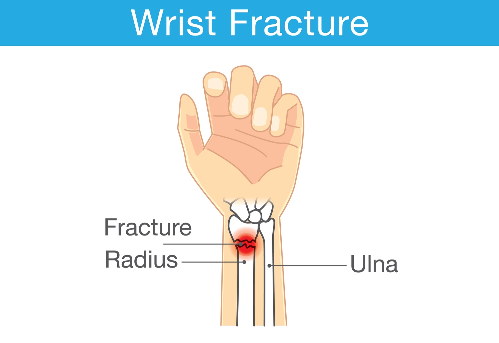
What Is a Distal Radius Fracture?
The radius is the larger of the two forearm bones. Its distal end forms part of the wrist joint. A distal radius fracture (DRF) is a break in this region, typically within 2 cm of the wrist joint.
DRFs account for approximately 17% of all fractures seen in emergency settings, ranging from simple undisplaced breaks treated with a cast to complex multi-fragment intra-articular injuries requiring sophisticated surgical fixation.
Most Common Fracture
DRF accounts for ~1 in 6 emergency room fractures
Bimodal Distribution
Most common in children 10-14 and adults over 60
Main Mechanism
Fall on outstretched hand (FOOSH injury)
Full Recovery Possible
With timely and appropriate specialist treatment
Common Causes of Distal Radius Fractures

Falls on Outstretched Hand
The most common mechanism — instinctively extending the hand to break a fall concentrates force at the distal radius, causing fracture.

Sports Injuries
Skiing, skateboarding, cycling, and contact sports carry high risk of wrist fractures particularly in younger and more active patients.

Osteoporosis in Elderly
Reduced bone density in older adults makes the distal radius vulnerable even to minor falls from standing height.
Symptoms to Watch For
Any suspicion of a wrist fracture after an injury should prompt immediate medical evaluation.
Immediate Pain
Sharp, severe pain at the wrist worsening with movement
Swelling
Rapid swelling around the wrist within minutes of injury
Bruising
Discolouration spreading over the wrist and hand
Deformity
“Dinner fork” appearance — wrist looks bent or misshapen
Tenderness
Point tenderness on pressing the distal radius bone
Loss of Function
Inability to move the wrist or grip objects properly
Seek Immediate Attention If:
You notice numbness, tingling, or weakness in the fingers — this may indicate nerve involvement requiring urgent care.

Types of Distal Radius Fractures
Classified by pattern, displacement, and involvement of the wrist joint surface.
Colles’ Fracture
The most common type — distal fragment displaces dorsally, creating the classic “dinner fork” deformity. Usually from a fall on an outstretched hand.
Smith’s Fracture
Reverse Colles’ — distal fragment displaces volarly. Less common; typically from a direct blow or fall on a flexed wrist.
Barton’s Fracture
Oblique fracture involving the rim of the distal radius with subluxation of the wrist joint. Often requires surgical intervention.
Chauffeur’s Fracture
Isolated fracture of the radial styloid process, often associated with wrist ligament injuries that require specialist assessment.
Intra-Articular Fracture
Fracture extends into the wrist joint surface. Surgical restoration of joint congruity is often required to prevent arthritis.
Comminuted Fracture
Bone shattered into multiple fragments. Most common after high-energy trauma or in osteoporotic bone. Usually requires surgical fixation.
Who Is at Risk?
Osteoporosis
Significantly reduces bone strength; even minor falls can cause fractures in affected individuals.
Age Over 60
Bone density decreases with age; postmenopausal women are especially vulnerable to fragility fractures.
Female Sex
Women have significantly higher lifetime fracture risk due to hormonal changes affecting bone density.
High-Risk Sports
Skiing, snowboarding, skateboarding, gymnastics — activities with high fall risk in younger patients.
Previous Fractures
A prior fragility fracture indicates compromised bone health and increased future fracture risk.
Low Calcium / Vitamin D
Nutritional deficiency weakens bones, increasing susceptibility to fractures from minimal trauma.
How Is It Diagnosed?
Clinical Assessment
History of injury mechanism, physical examination for deformity, swelling, and neurovascular status.
Plain X-Rays
AP and lateral wrist views — confirms fracture, assesses displacement, angulation, and shortening.
CT Scan
Required for complex intra-articular fractures to assess joint surface involvement and guide surgery.
MRI (If Indicated)
Used when soft tissue, ligament, or TFCC injuries are also suspected alongside the fracture.
Bone Density Scan
DEXA scan recommended for older patients to assess for underlying osteoporosis and guide long-term management.
Distal Radius Fracture Treatment
From conservative casting to precision surgical fixation — comprehensive treatment options tailored to your fracture type and lifestyle.
Non-Surgical Treatment
Appropriate for fractures that are undisplaced, minimally displaced, stable, and not significantly involving the joint surface.
Closed Reduction & Casting
If displaced, the fracture is reduced under anaesthesia without a surgical incision, then held in a cast with regular X-ray monitoring to ensure maintained position.
- Plaster or fibreglass cast for 6–8 weeks
- Repeat X-rays at 1, 2, and 4 weeks
- Finger exercises to prevent stiffness
- Elevation to reduce swelling
- NSAIDs and analgesics for pain relief
- Physiotherapy after cast removal
Who Qualifies for Non-Surgical Treatment?
Stable undisplaced fractures; minimally displaced fractures that can be reduced and held; elderly patients with lower functional demands; patients unfit for surgery.
Surgical Treatment Options
Surgery is recommended when fractures are significantly displaced, unstable, involve the joint surface, or cannot be controlled with a cast alone.
1. Open Reduction Internal Fixation (ORIF)
Gold standard for most displaced distal radius fractures. A volar locking plate is placed through a small incision on the palm side, providing rigid fixation allowing early wrist movement in physiotherapy.
2. Percutaneous K-wire Fixation
Small Kirschner wires inserted through the skin hold fracture fragments. Suitable for select fractures with good bone quality. Wires are usually removed at 4–6 weeks.
3. External Fixation
A frame attached outside the skin using pins above and below the fracture. Used for highly comminuted fractures or in cases with severe accompanying soft tissue injury.
Recovery & Rehabilitation Timeline
A structured, phased approach to recovery maximizes functional outcomes and return to normal activities.
Immediate Phase
Immobilization, swelling control, elevation, pain management, and finger exercises to maintain circulation.
Protected Mobilization
Gentle wrist range-of-motion exercises begin. Physiotherapy to restore joint movement and reduce stiffness.
Active Rehabilitation
Progressive strengthening, grip training, restoration of normal wrist mechanics, and return to light work.
Functional Recovery
Full return to daily activities, work, and most sports with advanced strengthening and sport-specific training.
Complete Recovery
Full bone healing confirmed on X-ray. Return to all high-demand activities and long-term outcome assessment.
Post-Treatment Care & When to See a Specialist
Signs of Good Healing
- Decreasing pain over weeks
- Reducing swelling after 1–2 weeks
- Progressively improving range of motion
- Normal X-ray findings at follow-up
Return Immediately If:
- Increasing or severe persistent pain
- Numbness or tingling in fingers
- Wound discharge post-surgery
- Fever or redness around wound
Specialist Review Schedule
- 1WFirst post-treatment review
- 2WRepeat X-ray to check alignment
- 6WCast removal (non-surgical)
- 3MFunctional assessment review
- 6MFinal outcome review
Dr. Vikas Gupta
Senior Consultant — Hand, Wrist & Shoulder Surgery | Founder, Hand2Shoulder Clinic
About Dr. Vikas Gupta
Dr. Vikas Gupta is one of India’s foremost specialists in hand, wrist, and shoulder surgery, with a distinguished career spanning over two decades. He completed his MS in Orthopaedics from a prestigious institution and underwent advanced fellowship training in Hand & Microsurgery internationally.
He founded the Hand2Shoulder Clinic to bring subspecialty-level, evidence-based care to patients suffering from upper limb conditions including distal radius fractures, carpal tunnel syndrome, tendon injuries, and shoulder disorders.
Dr. Gupta’s surgical expertise includes complex wrist fracture fixation using volar locking plate systems, arthroscopic wrist procedures, tendon repairs, nerve decompression, and shoulder reconstruction — all focused on restoring full function and minimizing recovery time.
Areas of Specialization
Distal Radius Fractures
Complex Wrist Fractures
Carpal Tunnel Syndrome
Tendon Repairs & Transfers
Wrist Arthroscopy
Shoulder Surgery
Expertise in Hand, Wrist & Shoulder Care
Advanced Surgical Techniques
Proficiency in volar locking plate fixation, arthroscopic wrist surgery, microvascular procedures, and complex reconstructive operations bringing international-level care to patients in India.

Patient Education & Communication
Known for clear, compassionate communication — every patient fully understands their diagnosis, treatment options, and recovery expectations before any decision is made.

Comprehensive Rehabilitation
Close collaboration with specialist physiotherapists to deliver outcome-focused recovery programmes, ensuring every patient achieves maximum functional restoration.
Distal Radius Fracture Articles
Expert-written educational content on wrist fractures, treatment, and recovery by Dr. Vikas Gupta and the Hand2Shoulder Clinic team.


Book a Consultation
Send us your details and Dr. Vikas Gupta’s team will respond within 24 hours to schedule your appointment.
Enquiry Form
All fields marked * are required. We respond within 24 hours.
Your information is kept strictly confidential and used only to respond to your enquiry.